Crystal Digital PCR™ Using EvaGreen®
EvaGreen®-based quantification in Crystal Digital PCR™
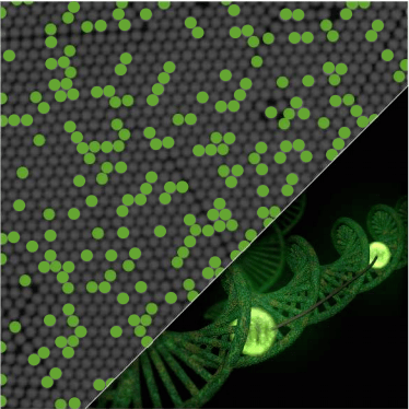
EvaGreen is a non-mutagenic, non-cytotoxic DNA-binding dye compatible with Crystal Digital PCR.
EvaGreen, which is non-fluorescent when free in solution, becomes strongly fluorescent upon binding to double-stranded DNA (dsDNA) in a sequence-independent manner. The signal generated can readily be detected using the same filters as those used for fluorescein.

Coupled with Crystal Digital PCR, EvaGreen thus enables absolute quantification of a target by using a simple primer pair. EvaGreen-based assays using our Sapphire chip yield reliable quantification down to 0.2 copies/ microliter.
Absolute quantification of genomic DNA using EvaGreen. A serial dilution of non-fragmented human genomic DNA, ranging from 1 000 to 0.2 copies per microliter was quantified by Crystal Digital PCR using 1.5X EvaGreen, 0.75X PerfecTa® UNG Toughmix, and oligonucleotides amplifying a 104 bp fragment from the EGFR gene. Reproducible and accurate quantification was obtained for the three replicates (R2= 0.9977). No positive droplets were observed in the non-template control wells.
Setting up EvaGreen® experiments
When setting up an experiment using EvaGreen, it is important to remember that high concentrations of any dsDNA initially present in the PCR mix will lead to higher basal fluorescence. Moreover, although EvaGreen has higher affinity for dsDNA, the dye will also be able to bind with lower affinity to any oligonucleotides present, thus also contributing to basal fluorescence.
Depending on the type of DNA that you want to quantify, and the intrinsic properties of your assay, you may need to assess the practical dynamic range.

Scatterplots from an EvaGreen experiment. A serial dilution of non-fragmented human genomic DNA, ranging from 1000 to 0.2 copies per microliter was quantified by Crystal Digital PCR using EvaGreen, 0.75X PerfeCT® UNG Toughmix, and oligonucleotides amplifying a 104 bp fragment from the EGFR gene.
Negative droplet populations are represented in grey, while positive droplet populations are represented in blue.
RFU: Relative Fluorescence Units